Tumor


Basics and background
The outer part of the cerebral hemispheres is formed by neocortex, with many gyri and sulci maximizing the brain surface. The sulci that appear first in development, the primary sulci, have a constant pattern, whereas the secundary and tertiary sulci show more interindividual variation.
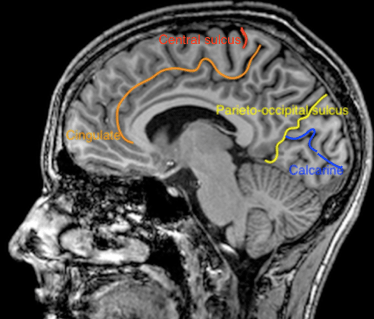
Figure 1. Primary sulci on parasagittal T1 weighted MRI
The brain is not made up of repeating units. The highly specialised functions vary depending on the anatomical location, e.g. the homunculi on the sensory and motor cortices as described by Wilder Penfield. The clinical presentation depends largely on the location of the pathology and is very different in case of a left frontal lobe lesion versus right occipital lobe pathology. (In contrast to the clinical presentation of left upper lobe versus right lower lobe lung disease.)
Every mm2 of cortical surface contains about 50,000-85,000 neurons supported by even larger numbers of glial cells. Microscopically, the neurons in the neocortex are arranged in six layers, that are numbered with Roman numerals from the outside to the inside. Each layer contains different types of neurons. The thickness and ratio of the layers varies per brain region.
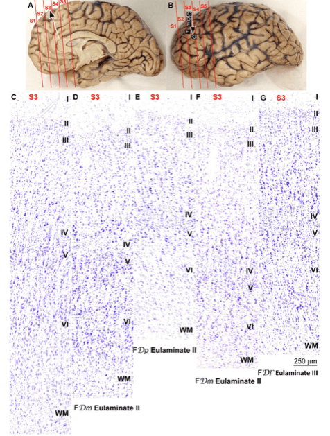
Figure 2. The 6 lamina of the human neocortex
From: Garcia-Cabezas MA, Hacker JL, Zikopoulos B A Protocol for Cortical Type Analysis of the Human Neocortex Applied on Histological Samples, the Atlas of Von Economo and Koskinas, and Magnetic Resonance Imaging Front Neuroanat 2020; 14: 576015 doi: 10.3389/fnana.2020.576015
When looking at the microscopy of human cortex with an untrained eye, it is difficult to see the separate layers. But in the work by Ramon y Cajal the difference between the pyramidal, polymorphic and granular cells is beautifully drawn, clarifying the distinctive layers of the cortex.
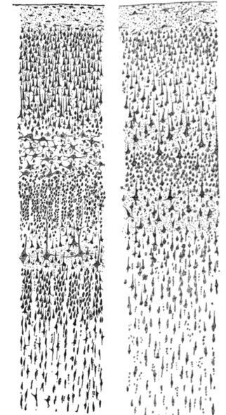
Figure 3. Drawings of the visual (left) and motor cortex (right) where the cell bodies (cytoplasm) of the neurons have been (Nissl) stained – by Santiago Ramon y Cajal. Originally published in 1899.
The majority of cells in the neocortex are excitatory glutamatergic neurons which arise from the ventricular zone of the telencephalon and migrate radially in waves to the surface. The first neurons form the outermost marginal layer (layer I) and prevent the next waves to overmigrate. Then the neurons from layer VI, V and IV migrate and finally the neurons from layer III and II (3-10).
At first this inside-out might seem unlogical because layer II neurons have to pass the previously migrated neurons. When you realise that layer II and III have mainly corticocortical connections and layer IV, V and VI connect to thalamus and brain stem, it makes sense: It is easier if the neurons that connect to the deeper structures are located on the inside (IV-VI), and it is not handy for neurons that communicate with other cortical regions (II-III) to migrate before these other cortical regions have been established.
The inhibitory GABAergic neurons migrate tangentially from the ganglionic eminence to their final location in the cortex, surrounded and outnumbered by glutamate neurons.
In the infratentorial cerebellum the opposite happens: GABAergic Purkinje cells arise from the ventricular layer and migrate radially and glutamatergic neurons from the rhombic lips migrate tangentially.
An increase in neuronal activity in a cortical region leads to an increase in local blood flow. fMRI is based on this neurovascular coupling and gives some insight in brain activity.
