Congenital/developmental
Metabolic
Tumor
Vascular


Basics and background
In the nineteen-sixties American neuroscientist Paul MacLean came up with an oversimplified and therefore practical and popular model of the brain. MacLean discerned 3 parts: the brainstem and basal ganglia, the limbic system and the neocortex.
1. The brainstem and basal ganglia were called the “reptilian brain”, involved in instinctual responses.
3. The neocortex or “human brain” houses all the higher brain functions such as language, mathematics, abstractian and thoughts. This makes up (only) half of the human brain volume.
2. The limbic system consists of the parts at the border of the brain stem and neocortex in the cerebral hemispheres. The limbic system is involved in emotion and memory and was called the “mammal brain” in the triune brain model.

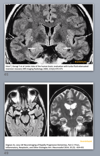
Figure 1. Coronal FLAIR image near the level of Monro’s foramen. The limbic structures (2) have a slightly higher signal intensity than the neocortex (3) because they are phylogenetically older and have a higher water content. Standard FLAIR images are not reliable for detecting abnormalities in the posterior fossa including the brain stem (1) and thalami.
The term “limbic system” is derived from the Latin word for border, “limbus”. As a teenager I liked Greek and Latin. In ancient Greece people wore a cloak that was actually a draped rectangular blanket kept in place by a pin. In men it was knee length and in women it reached to the ankle. More fortunate women had a border on their tunic or scarf , called parufė (woven-in) in Greek and limbus in Latin. In Aeneid Vergil describes limbus as a wavy border; you can imagine the hem of a dress moving when walking. Knowing that, for me “limbus” is not a static boundary.
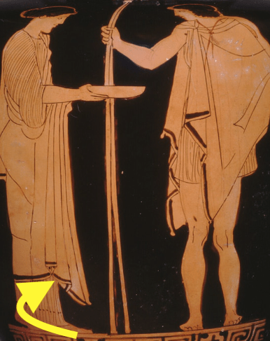
Figure 2. The man and woman wearing different garments, both with a limbus
Greek amphora from 455-445 B.C. – Chicago Art Institute http://www.artic.edu
The core structures of the limbic system are the hippocampus and amygdala, both located in the mesial temporal lobe. The parahippocampal gyrus, uncus, hypothalamus and cingulate gyrus are also part of the limbic system. The ventral tegmental area (VTA) in the midbrain and olfactory system are undeniably closely related and sometimes included in the limbic system. The “limbic system” is not a strictly defined anatomic system, but a functional concept.
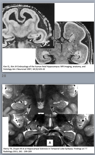
The hippocampus is the phylogenetically oldest part of brain cortex, consisting of only 3 layers and termed archicortex or archipallium. It consists of two interlocking gyri, the dentate gyrus and Ammon’s horn, and the subiculum. The hippocampus plays an important role in learning and memory, and also influences the hypothalamus. Precursor cells in the dentate gyrus keep forming new neurons (neurogenesis) during the entire human life, playing a role in the synaptic plasticity for learning and memory (15). The neighbouring parahippocampal gyrus and olfactory system are a little bit younger and are called paleocortex or paleopallium, consisting of 4 or 5 layers.

Figure 3. Hippocampus and amygdala on coronal IR image from the temporal lobe
At first sight the amygdala might resemble cortex, because it is gray matter located on the inner mesial surface of the temporal lobe. The amygdala is not cortex, but a group of about ten nuclei with specialised functions in fear, vigilance, emotion and memory (16).
Functionally the basolateral nuclei are often studied as an entity and these nuclei are especially sensitive to stress. The different basolateral nuclei all project to the central nucleus, the major output from the amygdaloid complex. The central amygdala connects to the bed nucleus of the stria terminalis (the gray matter on coronal MRI under the lateral ventricles that you always see but never notice) and several brain stem areas, such as the VTA in the midbrain, periaqueductal gray matter and hypothalamus. On coronal MR images you can see that the central amygdala is contiguous with the lentiform nucleus.
In contrast to the neocortex with glutamate as main neurotransmitter, the main neurotransmitter of the paleocortex is GABA and of the archicortex/hippocampus acetylcholine. The latter is also abundant in striate and thalami, although at some locations acetylcholine might function more as a neuromodulator than as an excitatory neurotransmitter.
*Note on figure 1: Standard FLAIR images are not reliable for detecting abnormalities in the posterior fossa and thalami.
The perceptibility of a lesion on MRI depends on the difference between the signal intensity of the lesion and the surrounding tissue (contrast) divided by the background noise.
The tissue in the brain stem is more heterogenous (gray and white matter intermixed) than supratentorial. Inflow of CSF and blood negatively influences the image quality of standard FLAIR images in the posterior fossa. By lengthening the inversion time and/or by performing 3D FLAIR, the visibility of infratentorial lesions can be improved.
