Inflammatory and infectious
Tumor


Basics and background
The brain stem and cerebellum are located in the posterior fossa. On imaging only a few structures are visible in the brain stem. The radiological location of brain stem lesions is mainly done by extrapolating histological knowledge and integrating it with surface morphology and relation to the visible structures e.g. red nucleus or aqueduct. A good clinician can locate brain stem pathology much better than current imaging studies.
The neurotransmitters serotonin, acetylcholine, noradrenaline and dopamine are distributed from the brain stem to the cortex.
Historically the divisions of the brain stem are based on the morphology. The middle part connecting to the cerebellum was named pons, with a “pregnant belly” on sagittal images. The upper part “for the eye movements” is the midbrain and the part below the pons is the medulla oblongata.
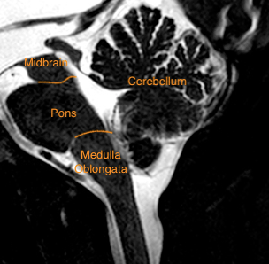
Figure 1 Brain stem divisions and cerebellum on sagittal T2 weighted image of the posterior fossa
In current medicine and with advancing knowledge we are moving more and more towards molecular based classifications of congenital malformations, tumors and other diseases. We are not there yet, but when discussing different pathologies in the vlogs, I will try to integrate as much as possible existing knowledge.
Luckily, animal models are more extrapolable in the brain stem than in the neocortex, because the brain stem is a highly conserved system. Referring to the article on which figure 2 is based, the gene expression during brain stem development in chicken embryos mirrors that of mouse embryos, evolutionary 300 million years separated. The advanced molecular knowledge of the brain stem brought to light that the 3-part morphological classification of the brain stem does not match the cell origins nor concurs with functional concepts.
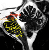
Figure 2 Divisions of the brain stem based on gene patterns and molecular expression
Based on: Watson C, Bartholomaeus C and Puelles L. Time for Radical Changes in Brain Stem Nomenclature—Applying the Lessons From Developmental Gene Patterns Front Neuroanat 2019; 13 :10 doi.org/10.3389/fnana.2019.00010
When it comes to gene expression the midbrain has more in common with the diencephalon and cerebral hemispheres (Otx2) than with the hindbrain (Gbx2). So in the future the midbrain might be included with supratentorial structures. The cephalic flexure bending the neural tube compresses the midbrain on the anterior side. On sagittal images the midbrain is therefore triangular and not parallel bounded. In several other species the dopaminergic cells are found low in the diencephalon, but in humans the dopaminergic region is “relocated” to the upper midbrain. Near the midbrain-hindbrain boundary the dopaminergic neurons have a very close anatomical relationship with the upper/rostral serotonergic neurons of the raphe nuclei (21).
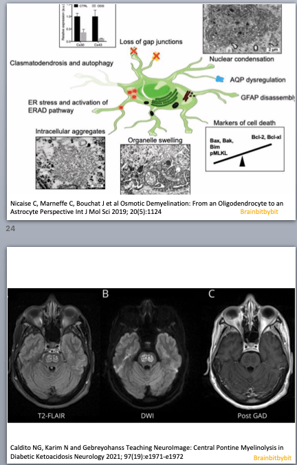
In the hindbrain or rhombencephalon, consisting of pons and medulla oblongata, vital functions are interdispersed with cranial nerve nuclei and ascending and descending tracts. Because of intermixed gray and white matter the pons is especially vulnerable to osmotic demyelination (45). The diverse neurological and psychiatric symptoms observed in patients with pontine osmotic demyelination reflect the different and divergent functions of this region. The brain’s main noradrenaline synthesis site is located in the dorsal pons, the locus coeruleus. From the rostral to the caudal hindbrain, raphe nuclei along the midline contain the majority of serotonergic neurons. The medulla oblongata or bulbus, houses the cardiovascular and respiratory center.
In gestational week 5-6 the cephalic, pontine and cervical flexure appear in the neural tube, displacing the alar plates laterally. The laterally displaced alar plates line a diamond (rhomboid) shaped membranous area and form the rhombic lips, subdivided in rhombomeres. From these rhombic lips, more specifically the rostral rhombomere r1, the cerebellar hemispheres are formed covering the upper part of the rhomboid membranous area, “closing” from superolateral like the curtains at the opera. The cerebellum has two germinal matrices: glutamatergic cells from the rhombic lips, and GABAergic Purkinje cells from the ventricular zone (23).
See also limbic system (end of page) – technical note on FLAIR: Standard FLAIR images are not reliable in the posterior fossa.
