Tumor


Basics and background
Basal ganglia and thalami
The basal ganglia are a group of nuclei located caudally in the prosencephalon. They consist of the caudate nucleus along the lateral ventricle and the lentiform nucleus which has an outer telencephalic part named putamen and an inner diencephalic part named globus pallidus or pallidum. The basal ganglia are best known for their function in fine tuning movement and motor control having feedback loops with the cortex (21-22, 54).

Figure 1. Upper row – coronal and sagittal drawing of secondary brain vesicles in the 6th and 7th gestational week From: https://embryology.med.unsw.edu.au
Bottom row – rotated (same) sagittal drawing and sagittal T1 weighted MR image of adult brain showing the caudate (Ca), putamen (Pu), globus pallidus (Gpa) and thalamus (Thal). The red arrow indicates the posterior and inferior growth of the caudate over the diencephalon.
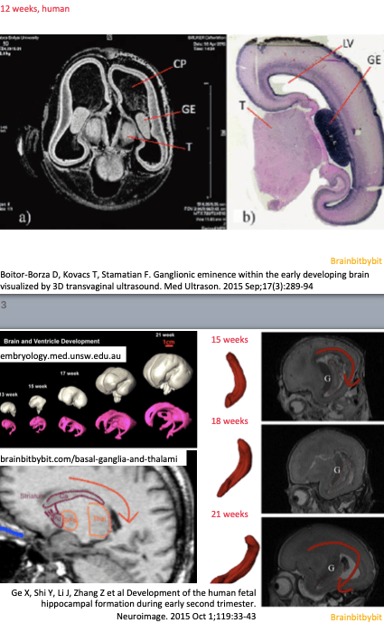
The caudate and putamen originate from the same structure which is named corpus striatum (in anticipation) and derived from the medial and lateral ganglionic eminences on the mesial, inner sides of the telencephalon. Between the 6th and 7th gestational week the telencephalon expands over the dorsal diencephalon/future thalamus, resulting in the C-shape of the caudate nuclei (and lateral ventricles). The ingrowing anterior limb of the internal capsule incompletely divides the corpus striatum into a medial caudate and lateral putamen and gives the striated appearance.
In the neocortex the GABAergic neurons originate from the ganglionic eminence and migrate tangentially mingling with the glutamatergic neurons from the ventricular zone. The main output neurotransmitter of the basal ganglia, which are derived of the ganglionic eminence, is also GABA. Acetylcholine is another abundant neurotransmitter in the basal ganglia, and thalami.
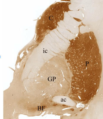
Figure 2. Coronal section of the brain of a 66-year old healthy female at the level of the anterior commissure (AC) showing part of the basal ganglia, stained for acetylcholine-esterase. Bridges of persistent gray matter in the internal capsule are the cause of the striate’s striped appearance. The part of the pallidum under the anterior commissure is called the ventral pallidum.
From: Jarret P, Easton A, Rockwood K et al Evidence for Cholinergic Dysfunction in Autosomal Dominant Kufs Disease Can J Neurol Sci 2018; 45(2): 150-157 doi: 10.1017/cjn.2017.261
The thalami are paired gray matter structures dorsal in the diencephalon on each side of the third ventricle. All the ascending fibers from the spinal cord and brain stem “switch” in different thalamic nuclei, and thalamocortical fibers project to different cortical areas. The thalamic nuclei are divided in an anterior, dorsomedial and (phylogenetically youngest) lateral group. On the dorsal side of the thalamus are two geniculate bodies: the medial geniculate body for auditory information and the lateral geniculate body as part of the visual pathway. The thalamus is part of the motor control feedback system with the basal ganglia, receiving input from the internal globus pallidus and projecting to the motor cortex. The thalamus is not just a relay station, but works like a good secretary estimating all the incoming information and sending it to the correct cortical region. In addition the thalamus has an important role in consciousness and the sleep-wake cycle. The interthalamic adhesion, which is formed by fusion, connects the thalami and still has a lot of ambiguousness.
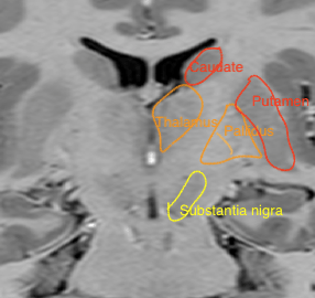
Figure 3. Coronal inversion recovery image at the level of the foramen of Monro. The difference in signal intensity between the diencephalic (thalamus and pallidus) and telencephalic structures (caudate nucleus and putamen) is visible. The thalamus is located as cranial as the insular cortex.
Because of their role in the motor control system, the subthalamic nucleus and the substantia nigra rostrally in the midbrain are often included when referring to the basal ganglia.
Centrencephalon
I want to add a bit that is more about the mind than the brain: the centrencephalon, a term first used by Wilder Penfield in the 1950s.
Penfield was a neurosurgeon who performed awake craniotomy in epilepsy patients using electrical stimulation and greatly improved our knowledge of the brain. He mapped the homunculus on the motor cortex and discovered together with Brenda Milner the role of the hippocampus in memory.
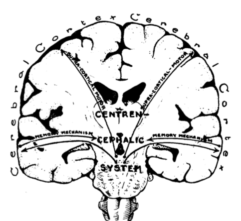
Figure 4. Drawing of the centrencephalic system
From: Penfield W, Jasper HH Epilepsy and the functional anatomy of the human brain (book – Little, Brown and company 1954)
He came up with the concept of a Centrencephalic Integrating System (CIS) in which the centrencephalon, consisting of thalami and higher brain stem, integrates all information from the cortex of both hemispheres. The CIS was considered the substrate of consciousness and mind: Like the motor cortex consults the basal ganglia to improve movement, the frontal cortex consults the basal network in reward detection (a.o. ventral striatum and ventral pallidum), motivation and cognition. Our knowledge of the motor network is greater than our comprehension of human behaviour because the output is easier to measure, not because movement is more important than thoughts or imagination.
There is a quote from Wilder Penfield, which really appealed to me, on the facade of the Montreal Neurological Institute: “The problem of neurology is to understand man himself”.
